Cross Section Of A Bone Diagram ~ Bone marrow cell populations. (a) Cross section of tubular bone showing... | Download Scientific .... The outside of a bone is covered in a thin layer of dense irregular connective tissue called the periosteum. Looking at a bone in cross section, there are several distinct layered regions that make up a bone. Diagram of channel cross section leaf cross section diagram label worksheets. The cross section of a rectangular pyramid is a rectangle. Two types of bone tissues in cross section of a long bone :
Labelled diagram of hip bone wiring diagram t1. Explaned distal and proximal epiphysis. Spongy bone diagram schematic diagram. Some descriptions for confusing partsomit number 13 in the picture. There are two ways to study bone histology.
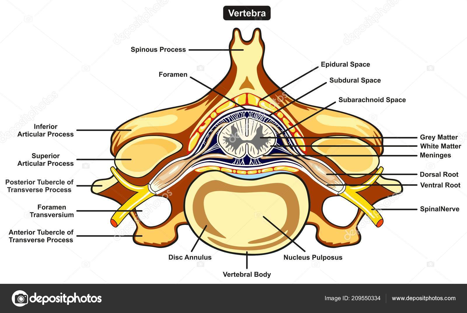
Human body labeled | Labeled Vertebra Cross Section Human Body Anatomy Infographic Diagram ... from st4.depositphotos.com In the last decade, considerable technological improvements have been made to repair damaged bones and tissue, such as bone cross sections with implants for microscopic examinations. We don't draw the rest of the object, just the shape made when you cut through. Labelled diagram of hip bone wiring diagram t1. Jump to navigation jump to search. Cross sections are usually parallel to the base like above, but can be in any direction. Diagram with articular cartilage, marrow, spongy bone, medullary cavity, endosteum, diaphysis, and periosteum. Bone contains a relatively small number of cells entrenched in a matrix of collagen fibers that provide a surface for inorganic salt crystals to adhere. Cross section of a plant leaf diagram.
As shown in figure 2.
Dry bone is cut and polished before mounting on a slide. We can see there are two layers of compact bone here. Bone marrow is the soft, highly vascular and flexible connective tissue within bone cavities which serve as the primary site of new blood cell production or bone marrow is the primary source of pluripotent stem cells that give rise to all hemopoietic cells (blood cells) including lymphocytes. Whereas a long bone has only one layer of compact bone (see fig 1). 21.09.2020 · cross section of the leg through the soleus muscle: Vector illustration scheme of bone cross section. Two types of bone tissues in cross section of a long bone : Find the perfect bone diagram stock illustrations from getty images. The answer lies in the properties of a third. The initial step involves the development of a cartilage model, which has the rough shape of the bone being formed. Diagram of channel cross section leaf cross section diagram label worksheets. In the middle of the shaft is the. Bone is found in the shafts of long bone and consists of various cylindrical units named as haversian system 47.
Internal structure of the dicotyledonous stem by openstax. A cross section of a human long bone. Medically reviewed by the healthline medical network — written by the healthline editorial team — updated on january 20, 2018. Generally speaking, it is very easy to recognize a cross section through the leg, mostly due to the tibia. Two types of bone tissues in cross section of a long bone :
Cross-section of the Long Bone from wcs.smartdraw.com A long bone has two main regions: Cross section of bone diagram. Diagram with articular cartilage, marrow, medullary cavity and periosteum. Bone cross section diagram ipad folio cases. As a part of the. In the last decade, considerable technological improvements have been made to repair damaged bones and tissue, such as bone cross sections with implants for microscopic examinations. Vector illustration scheme of bone cross section. It seems confusing and misleading.
21.09.2020 · cross section of the leg through the soleus muscle:
The periosteum contains many strong collagen fibers that are used to firmly anchor. In the last decade, considerable technological improvements have been made to repair damaged bones and tissue, such as bone cross sections with implants for microscopic examinations. Medically reviewed by the healthline medical network — written by the healthline editorial team — updated on january 20, 2018. Labelled diagram of hip bone wiring diagram t1. The answer lies in the properties of a third. Spongy bone is composed of trabeculae that contain the osteocytes. Internal structure of the dicotyledonous stem by openstax. Cross section of the long bone. Whereas a long bone has only one layer of compact bone (see fig 1). Skin anatomy diagram description illustration skin stock. I am not an expert on this subject, so i was wondering if i don't like way you've shown the cartilage. The initial step involves the development of a cartilage model, which has the rough shape of the bone being formed. Dry bone is cut and polished before mounting on a slide.
Diagram with articular cartilage, marrow, spongy bone, medullary cavity, endosteum, diaphysis, and periosteum. I am not an expert on this subject, so i was wondering if i don't like way you've shown the cartilage. Bone cross section diagram ipad folio cases. A cross section of a human long bone. There are two ways to study bone histology.
Chapter 6 Bones and Cartilage - Biology 4 Human AnatomyProfessor Julie GallagherBarstow ... from biology4bcc.weebly.com Cross section of bone diagram. Diagram with articular cartilage, marrow, spongy bone, medullary cavity, endosteum, diaphysis, and periosteum. Bone decalcification is the removal of the mineral component using an acid, leaving the bone soft and easy to cut. There are two ways to study bone histology. A long bone has two main regions: A cross section of a human long bone. It seems confusing and misleading. Whereas a long bone has only one layer of compact bone (see fig 1).
They build the entire picture, improve your understanding, consolidate the information and facilitate recall. The hip bone ilium ischium pubis teachmeanatomy. Spongy bone diagram schematic diagram. Diagram with articular cartilage, marrow, medullary cavity and periosteum. Cross sections are usually parallel to the base like above, but can be in any direction. Later discussions in this chapter will show that bone is also dynamic in that its if osteoblasts and osteocytes are incapable of mitosis, then how are they replenished when old ones die? In the last decade, considerable technological improvements have been made to repair damaged bones and tissue, such as bone cross sections with implants for microscopic examinations. As a part of the. Labelled diagram of hip bone wiring diagram t1. Internal structure of the dicotyledonous stem by openstax. Fermur bone with labels and diagram. Classify each of the following terms as a projection (p) or a depression (c) identify one lamella on diagram a by using a bracket and label (the concentric ellae would be difficult to color without confusing other structures) lacunae. Dry bone is cut and polished before mounting on a slide.
This bone is located directly beneath the skin on the anterior aspect of the leg (top of the image) cross section of a bone. Fermur bone with labels and diagram.
Share :
Post a Comment
for "Cross Section Of A Bone Diagram ~ Bone marrow cell populations. (a) Cross section of tubular bone showing... | Download Scientific ..."


 Cross section of tubular bone showing... | Download Scientific ...)
Post a Comment for "Cross Section Of A Bone Diagram ~ Bone marrow cell populations. (a) Cross section of tubular bone showing... | Download Scientific ..."