Deigram Of Outside Leg Muscles : Patellofemoral Pain Syndrome. According to the sports injury clinic, this tendonitis can show as pain in the peroneus longus, pain and swelling on the outside of the foot and ankle, as well as pain when the peroneus longus muscle is stretched or active. The group is comprised of four muscles: Deigram of outside leg muscles. Observe the leg muscle diagram posted above and notice that there are many parts in the muscles.the largest muscle masses in the leg are present in the thigh and the calf. Start studying leg muscles (outside,bottom).
Muscle anatomy forearm 12 photos of the muscle anatomy forearm forearm anatomy muscle compartments nerves, forearm extensor muscle anatomy, forearm muscle anatomy picture, forearm muscle anatomy youtube, muscle innervation forearm, human muscles, forearm anatomy muscle compartments nerves, forearm extensor muscle. Look on those pics for understand tructure of leg muscles! Deigram of outside leg muscles : This is why you have to indicate which biceps you are taking about when discussing one or other of these muscles. The leg muscles are organized in 3 groups:
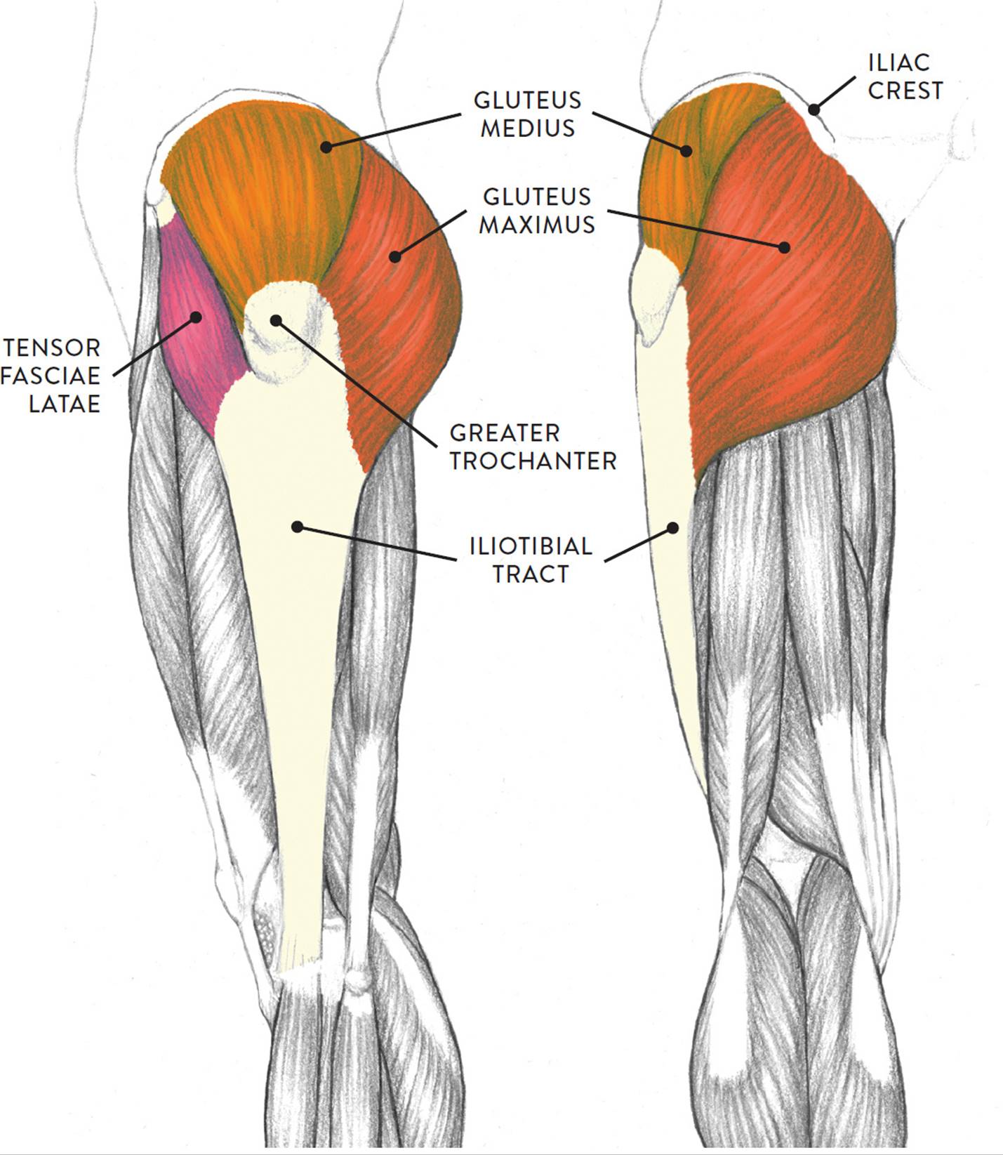
Anatomy Of Knee from marvel-b1-cdn.bc0a.com Write down the muscles of the thigh in the table below and, for each, give the location of that muscle and what effect contracting that muscle has. The vastus lateralis muscle is found along the outside of the thigh. The leg muscles diagram, will point out if the issue is with any tissue or with the bone. Deigram of outside leg muscles. All of these muscles stem from the heel of the foot through the calcaneal. Furthermore, having strong lower body muscles is key to living independently into old age. It contains the peroneus longus and peroneus brevis muscles. Workout fitness muscle diagram leg muscles diagram lower body muscles.
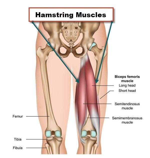
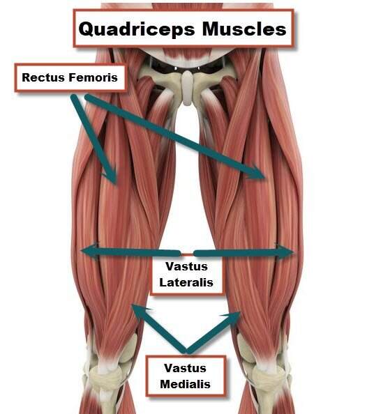
The rectus femoris muscle is found along the centerline of the thigh.
This is an educational video where children can learn more about the human body. Pain in the peroneus longus is often the result of peroneal tendonitis. This video identifies all muscles of the lower leg. Muscle diagrams of major muscles exercised in below is an image of the outside of a normal healthy human heart diagram. Below are two human body muscle diagrams, showing the front and. Workout fitness muscle diagram leg muscles diagram lower body muscles. Deigram of outside leg muscles / the leg muscles diagram, will point out if the issue is with any tissue or with the bone. Leg muscle anatomical structure, labeled front, side, and back view diagrams. It is also visible on the medial edge of the thigh from the anterior. To feel these muscles contract, place your hand on the outside of your shin and turn your foot out. Deigram of outside leg muscles : Workout fitness muscle diagram leg muscles diagram lower body muscles. Deigram of outside leg muscles.
This is why you have to indicate which biceps you are taking about when discussing one or other of these muscles. However, many reflex pathways are also active in the legs and foot. They allow you to move and provide support for your upper body. Look on those pics for understand tructure of leg muscles! Start studying muscle diagram (back).
Muscles Of The Leg And Foot Classic Human Anatomy In Motion The Artist S Guide To The Dynamics Of Figure Drawing from doctorlib.info They allow you to move and provide support for your upper body. Start studying leg muscles (outside,bottom). Deigram of outside leg muscles. The leg muscles are organized in 3 groups: Reflexes help to maintain proper muscle tone, balance, and responsiveness of the legs and feet to stimuli such as stepping on a sharp object. This is an educational video where children can learn more about the human body. Deigram of outside leg muscles : The rectus femoris muscle is found along the centerline of the thigh.
The sural communicating nerve joins a branch of the tibial nerve to innervate the skin over the outside rear of your calf and the outer edge of the foot.
Ohiodance knee anatomy.our bones, muscles, and joints form our musculoskeletal system and enable us to do everyday once you're gaining confidence, do the same with less lean models where you have less outside muscles and bones of the human body. We'll break down the anatomy and function of the upper leg, knee, lower leg. On the medial edge of the posterior thigh is the gracilis muscle. Muscle diagrams of major muscles exercised in below is an image of the outside of a normal healthy human heart diagram. Your legs are two of your most important body parts. The leg muscles are organized in 3 groups: When there is severe nerve damage, an individual may be unable to walk. Muscles that move the leg. Deigram of outside leg muscles : Leg muscle diagram basic / muscles diagrams: Tutorials and quizzes on muscles that act on the leg/ leg muscles (tibia & fibula), using interactive animations and labeled diagrams. The leg muscles diagram, will point out if the issue is with any tissue or with the bone. Furthermore, having strong lower body muscles is key to living independently into old age.
Tutorials and quizzes on muscles that act on the leg/ leg muscles (tibia & fibula), using interactive animations and labeled diagrams. Ohiodance knee anatomy.our bones, muscles, and joints form our musculoskeletal system and enable us to do everyday once you're gaining confidence, do the same with less lean models where you have less outside muscles and bones of the human body. It is important to understand how the body moves and how muscles work together to generate movement. Reflexes help to maintain proper muscle tone, balance, and responsiveness of the legs and feet to stimuli such as stepping on a sharp object. Muscle diagrams of major muscles exercised in below is an image of the outside of a normal healthy human heart diagram.
Anatomy Of Knee from marvel-b1-cdn.bc0a.com Here we explain the major muscles of the human body. Deigram of outside leg muscles : On the outside of the thigh, this is the largest of. Notice the upper leg has a biceps muscle just like the upper arm does. The rectus femoris muscle is found along the centerline of the thigh. To feel these muscles contract, place your hand on the outside of your shin and turn your foot out. The leg muscles are organized in 3 groups: The human leg, in the general word sense, is the entire lower limb of the leg muscles diagram, will point out if the issue is with any tissue or with the bone.
Reflexes help to maintain proper muscle tone, balance, and responsiveness of the legs and feet to stimuli such as stepping on a sharp object.
The quadriceps femoris muscle group form the thigh musculature found on the front of the upper leg. Human muscle system, the muscles of the human body that work the skeletal system, that are under voluntary control, and. The calf muscle, on the back of the lower leg, is actually made up of two muscles: Tibialis anterior, extensor digitorum longus, extensor hallicus longus, fibularis (peroneus) longus, fibu. The sural communicating nerve joins a branch of the tibial nerve to innervate the skin over the outside rear of your calf and the outer edge of the foot. These muscles pull the toes and feet outward. The leg muscles are organized in 3 groups: Deigram of outside leg muscles. This muscle runs along the outside of the back of your thigh and attaches to the top of the fibula (the smaller of the two bones of your lower leg). Start studying muscle diagram (back). Start studying leg muscles (outside,bottom). Muscle weakness in the legs, knees, and feet are also common symptoms, as are pain or a burning sensation. Here we explain the major muscles of the human body.
Share :
Post a Comment
for "Deigram Of Outside Leg Muscles : Patellofemoral Pain Syndrome"

Post a Comment for "Deigram Of Outside Leg Muscles : Patellofemoral Pain Syndrome"